Historically,
many Egyptologists focused primarily on the very visible aspects of
ancient Egyptian society, such as the pyramids, much to the bain of
those interested in more than just monumental architecture. From the
beginning of the scholarly study of Egypt's past there have been few
scholars who recognized the importance of the process of disease and
health on a population. With the turn of the century, new archaeological
discoveries, increased knowledge of Egyptian language and writing, and
the advent of more sophisticated medical techniques, new life was
breathed into the study of disease and health in the ancient Nile
Valley. It was this period that saw the academic study of Egyptian
disease segregated into three distinct categories.
The first is the study of medical Papyri. Early on it was recognized
that the textual material of the Dynastic Period pertaining to the
recognition and treatment of disease was extremely important for
understanding both the state of health as well as the concept of disease
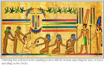
in ancient Egypt. The second is the study of the artistic
representation of disease in the Nile Valley. The Egyptian's
predilection to portrayl life in a relatively realistic manner offers an
excellent opportunity for the study of disease. The third, and perhaps
most obvious, is the study of human remains, both skeletal and soft
tissue, of ancient Egyptians. With the advent of increasingly
sophisticated medical techniques at the beginning of the 20th century,
as well as those complex medical techniques in use today, the analysis
of Egypt's veritable wealth of human remains provided a tremendous boost
to the study of the state of disease and health in the ancient Nile
Valley.
Medical Papyri
The Edwin Smith Papyrus
The Edwin
Smith Surgical Papyrus is, without a doubt, one if the most important
documents pertaining to medicine in the ancient Nile Valley. Placed on
sale by Mustafa Agha in 1862, the papyrus was purchased by Edwin Smith.
An American residing in Cairo, Smith has been described as an
adventurer, a money lender, and a dealer of antiquities.(Dawson and
Uphill: 1972). Smith has also been reputed as advising upon, and even
practicing, the forgery of antiquities.(Nunn 1996:26) Whatever his
personal composition, it is to his credit that he immediately recognized
the text for what it was and later carried out a tentative translation.
Upon his death in 1906, his daughter donated the papyrus in its
entirety to the New York Historical Society. The papyrus now resides in
the collections of the New York Academy of Sciences.
In 1930, James Henry Breasted, director of the Oriental Institute at the
University of Chicago, published the papyri with facsimile,
transcription, English translation, commentary, and introduction. The
volume was accompanied by medical notes prepared by Dr. Arno B.
Luckhardt. To date, the Breasted translation is the only one if its
kind.
The Edwin Smith papyrus is second in length only to the Ebers papyrus,
comprising seventeen pages (377 lines) on the recto and five pages (92
lines) on the verso. Both the recto and the verso are written with the
same hand in a style of Middle Egyptian dating.
The Ebers Papyrus
Like the
Edwin Smith Papyrus, the Ebers Papyrus was purchased in Luxor by Edwin
Smith in 1862. It is unclear from whom the papyrus was purchased, but it
was said to have been found between the legs of a mummy in the Assassif
district of the Theben necropolis.
The papyrus remained in the collection of Edwin Smith until at least
1869 when there appeared, in the catalog of an antiquities dealer, and
advertisement for "a large medical papyrus in the possession of Edwin
Smith, an American farmer of Luxor."(Breasted 1930) The Papyrus was
purchased in 1872 by the Egyptologist George Ebers, for who it is named.
In 1875, Ebers published a facsimile with an English-Latin vocabulary
and introduction.
The Ebers Papyrus comprises 110 pages, and is by far the most lengthy of
the medical papyri. It is dated by a passage on the verso to the 9th
year of the reign of Amenhotep I (c. 1534 B.C.E.), a date which is close
to the extant copy of the Edwin Smith Papyrus. However, one portion of
the papyrus suggests a much earlier origin. Paragraph 856a states that :
"the book of driving wekhedu from all the limbs of a man was found in
writings under the two feet of Anubis in Letopolis and was brought to
the majesty of the king of Upper and Lower Egypt Den."(Nunn 1996: 31)
The reference to the Lower Egyptian Den is a historic anachronism which
suggesting an origin closer to the First Dynasty (c. 3000 B.C.E.)
Unlike the Edwin Smith Papyrus, the Ebers Papyrus consists of a
collection of a myriad of different medical texts in a rather haphazard
order, a fact which explains the presence of the above mentioned
excerpt. The structure of the papyrus is organized by paragraph, each of
which are arranged into blocks addressing specific medical ailments.
Paragraphs 1-3 contain magical spells designed to protect from
supernatural intervention on diagnosis and treatment. They are
immediately followed by a large section on diseases of the stomach
(khet), with a concentration on intestinal parasites in paragraphs
50-85.(Bryan 1930:50) Skin diseases, with the remedies prescribed placed
in the three categories of irritative, exfoliative, and ulcerative, are
featured in paragraphs 90-95 and 104-118. Diseases of the anus,
included in a section of the digestive section, are covered in
paragraphs 132-164.(Ibid. 50) Up to paragraph 187, the papyrus follows a
relatively standardized format of listing prescriptions which are to
relieve medical ailments. However, the diseases themselves are often
more difficult to translate. Sometimes they take the form of
recognizable symptoms such as an obstruction, but often may be a
specific disease term such as wekhedu or aaa, the meaning of both of
which remain quite obscure.

Paragraphs
188-207 comprise "the book of the stomach," and show a marked change in
style to something which is closer to the Edwin Smith Papyrus.(Ibid.:
32) Only paragraph 188 has a title, though all of the paragraphs include
the phrase: "if you examine a man with a…," a characteristic which
denotes its similarity to the Edwin Smith Papyrus. From this point, a
declaration of the diagnosis, but no prognosis. After paragraph 207, the
text reverts to its original style, with a short treatise on the heart
(Paragraphs 208-241).
Paragraphs 242-247 contains remedies which are reputed to have been made
and used personally by various gods. Only in paragraph 247, contained
within the above mentioned section and relating to Isis' creation of a
remedy for an illness in Ra's head, is a specific diagnosis mentioned.
(Bryan 1930:45)
The following section continues with diseases of the head, but without
reference to use of remedies by the gods. Paragraph 250 continues a
famous passage concerning the treatment of migraines. The sequence is
interrupted in paragraph 251 with the focus placed on a drug rather than
an illness. Most likely an extract from pharmacopoeia, the paragraph
begins: "Knowledge of what is made from degem (most likely a ricinous
plant yielding a form of castor oil), as something found in ancient
writings and as something useful to man."(Nunn 1996: 33)
Paragraphs 261-283 are concerned with the regular flow of urine and are
followed by remedies "to cause the heart to receive bread."(Bryan
1930:80). Paragraphs 305-335 contain remedies for various forms of
coughs as well as the genew disease.
The remainder of the text goes on to discuss medical conditions
concerning hair (paragraphs 437-476), traumatic injuries such as burns
and flesh wounds (paragraphs 482-529), and diseases of the extremities
such as toes, fingers, and legs. Paragraphs 627-696 are concerned with
the relaxation or strengthening of the metu. The exact meaning of metu
is confusing and could be alternatively translated as either mean hollow
vessels or muscles tissue.(Ibid.:52) The papyrus continues by featuring
diseases of the tongue (paragraphs 697-704), dermatological conditions
(paragraphs 708-721), dental conditions (paragraphs 739-750), diseases
of the ear, nose, and throat (paragraphs 761-781), and gynecological
conditions (paragraphs 783-839)
Kahun Gynecological Papyrus
The Kahun
Papyrus was discovered by Flinders Petrie in April of 1889 at the Fayum
site of Lahun. The town itself flourished during the Middle Kingdom,
principally under the reign of Amenenhat II and his immediate successor.
The papyrus is dated to this period by a note on the recto which states
the date as being the 29th year of the reign of Amenenhat III (c. 1825
B.C.E.). The text was published in facsimile, with hieroglyphic
transcription and translation into English, by Griffith in 1898, and is
now housed in the University College London.
The gynecological text can be divided into thirty-four paragraphs, of
which the first seventeen have a common format.(Nunn 1996: 34) The first
seventeen start with a title and are followed by a brief description of
the symptoms, usually, though not always, having to do with the
reproductive organs.
The second section begins on the third page, and comprises eight
paragraphs which, because of both the state of the extant copy and the
language, are almost unintelligible. Despite this, there are several
paragraphs that have a sufficiently clear level of language as well as
being intact which can be understood. Paragraph 19 is concerned with the
recognition of who will give birth; paragraph 20 is concerned with the
fumigation procedure which causes conception to occur; and paragraphs
20-22 are concerned with contraception. Among those materials prescribed
for contraception are crocodile dung, 45ml of honey, and sour
milk.(Ibid:35)
The third section (paragraphs 26-32) is concerned with the testing for
pregnancy. Other methods include the placing of an onion bulb deep in
the patients flesh, with the positive outcome being determined by the
odor appearing to the patients nose.
The fourth and final section contains two paragraphs which do not fall
into any of the previous categories. The first prescribes treatment for
toothaches during pregnancy. The second describes what appears to be a
fistula between bladder and vagina with incontinence of urine "in an
irksome place."(Ibid. 35)
The Investigation of Disease Patterns Through Human Remains and Artistic Representations
Parasitic Diseases
Schistosomiasis (bilharziasis)
Of the
three main species of the platyhelminth worm Schistosoma, the most
important for Egypt are S. mansoni and S. haematobium. There is a
complex life cycle alternating between two hosts, humans and the fresh
water snail of the genus Bulinus. The infection is caught by humans who
come into contact with the free swimming worm which the snail releases
in the water. The worm penetrates the intact skin and enters the veins
of the human host. The main symptom of the presence of the parasite is
haematuria which results in serious anemia, loss of appetite, urinary
infection, and loss of resistance to other diseases. There may also be
interference with liver functions.
One of the finest archaeological examples for the existence of
schistosomiasis in ancient Egypt was the discovery of calcified ova in
the unembalmed 21st Dynasty mummy of Nakht. Upon medical examination,
the mummy not only exhibited a preserved tapeworm, but also ova of the
Schistosoma haematobium and displayed changes in the liver resulting
from a schistosomal infection.(Millat et al. 1980:79)
Bacterial and Viral Infections
Tuberculosis (Mycobacterium tuberculosis)
Ruffer
(1910) reported the presence of tuberculosis of the spine in Nesparehan,
a priest of Amun of the 21st Dynasty. This shows the typical features
of Pott's disease with collapse of thoracic vertebra, producing the
angular kyphosis (hump-back). A well known complication of Pott's
disease is the tuberculous suppuration moving downward under the psoas
major muscle, towards the right iliac fossa, forming a very large psoas
abscess.(Nunn 1996:64)
Ruffer's report has remained the best authenticated case of spinal
tuberculosis from ancient Egypt. All known possible cases, ranging from
the Predynastic to 21st Dynasty were reviewed by Morse, Brockwell, and
Ucko (1964) as well as by Buikstra, Baker, and Cook.(1993) These
included Predynastic specimens collected at Naqada by Petrie and Quibell
in 1895 as well as nine Nubian Specimens from the Royal College of
Surgeons of England. Both reviewers were in agreement that there was
very little doubt that tuberculosis was the cause of pathology in most,
but not all, cases. In some cases, it was not possible to exclude
compression fractures, osteomyelitis, or bone cysts as causes of death.
The numerous artistic representation of hump-backed individuals are
provocative but not conclusive. The three earliest examples are
undoubtedly of Predynastic origin. The first is a ceramic figurine
reported to have been found by Bedu in the Aswan district. It represents
an emaciated human with angular kyphosis of the thoracic spine
crouching in a clay vessel.(Schrumph-Pierron 1933) The second possible
Predynastic representation with spinal deformity indicative of
tuberculosis is a small standing ivory likeness of a human with arms
down at the sides of the body bent at the elbows. The head is modeled
with facial features carefully indicated. The figure is shown with a
protrusion of the back and on the chest.(Morse 1967: 261) The last
Predynastic example is a wooden statue contained within the Brussels
Museum. Described as a bearded male with intricate facial features, the
figure has a large rounded hunch-back and an angular projection of the
sternum.(Jonckheere 1948: 25)
As well, there are several historic Egyptian representations which
indicate the possibility of tuberculosis deformity. One of the most
suggestive, located in and Old Kingdom 4th Dynasty tomb, is of a bas
relief serving girl who exhibits localized angular kyphosis. A second
provocative example has its origin in the Middle Kingdom. A tomb
painting at Beni Hasan, the representation shows a gardener with a
localized angular deformity of the cervical-thoracic spine.(Morse 1967:
263)
Poliomyelitis
A viral
infection of the anterior horn cells of the spinal chord, the presence
of poliomyelitis can only be detected in those who survive its acute
stage. Mitchell (Sandison 1980:32) noted the shortening of the left leg,
which he interpreted as poliomyelitis, in the an early Egyptian mummy
from Deshasheh. The club foot of the Pharaoh Siptah as well as
deformities in the 12th Dynasty mummy of Khnumu-Nekht are probably the
most attributable cases of poliomyelitis.
An 18th or 19th Dynasty funerary staele shows the doorkeeper Roma with a
grossly wasted and shortened leg accompanied by an equinus deformity of
the foot. The exact nature of this deformity, however, is debated in
the medical community. Some favor the view that this is a case of
poliomyelitis contracted in childhood before the completion of skeletal
growth. The equinus deformity, then, would be a compensation allowing
Roma to walk on the shortened leg. Alternatively, the deformity could be
the result of a specific variety of club foot with a secondary wasting
and shortening of the leg.(Nunn 1996: 77)
Deformities
Dwarfism
Dasen
(1993) lists 207 known representations of dwarfism. Of the types
described, the majority are achondroplastic, a form resulting in a head
and trunk of normal size with shortened limbs. The statue of Seneb is
perhaps the most classic example. A tomb statue of the dwarf Seneb and
his family, all of normal size, goes a long way to indicate that dwarfs
were accepted members in Egyptian society. Other examples called
attention to by Ruffer (1911) include the 5th Dynasty statuette of
Chnoum-hotep from Saqqara, a Predynastic drawing of the "dwarf Zer" from
Abydos, and a 5th Dynasty drawing of a dwarf from the tomb of
Deshasheh.
Skeletal evidence, while not supporting the social status of dwarfs in
Egyptian society, does corroborate the presence of the deformity. Jones
(Brothwell 1967:432) described a fragmentary Predynastic skeleton from
the cemetery at Badari with a normal shaped cranium both in size in
shape. In contrast to this, however, the radii and ulna are short and
robust, a characteristic of achondroplasia. A second case outlined by
Jones (Ibid.:432) consisted of a Predynastic femur and tibia, both with
typical short shafts and relatively large articular ends.
References
Breasted, J.H. - The Edwin Smith Surgical Papyrus (University of Chicago Press: University of Chicago, 1930)
Brothwell,
D. - "Major Congenital Anomalies of the Skeleton," in Diseases in
Antiquity: A Survey of Disease, Injuries, and Surgery in Early
Populations (eds.) A.T. Sandison and D. Brothwell (Charles C. Thomas:
Springfield, 1967)
Bryan, P.W. - The Papyrus Ebers (Geoffrey Bles: London, 1930)
Buikstra,
J.E.; Baker, B.J.; Cook, D.C.- "What Disease Plagues the Ancient
Egyptians? A Century of Controversy Considered," In Biological
Anthropology and the Study of Ancient Egypt (eds.) W,V. Davies and R.
Walter (British Museum Press: London, 1993)
Dasen, V. - Dwarfs in Ancient Egypt and Greece (Clarendon Press: Oxford, 1993)
Dawson, W.R. and E.P. Uphill - Who Was Who in Egyptology (Egyptian Exploration Society: London, 1993)
Jonckheere, F. - "Le Bossu des Mussées Royaux D'Art et D'Histoire de Bruxelles," Chronique D'Égypt (45) 25, 1958.
Millet,
N.; Hart, G.; Reyman, T.; Zimerman, A.; Lewein, P. - "ROM I:
Mummification for the Common People," in Mummies, Disease, and Ancient
Cultures (eds.) Aiden and Eve Cockburn (Cambridge University Press:
Cambridge, 1980)
Morse,
D. - "Tuberculosis," in Diseases in Antiquity: A Survey of Diseases,
Injuries, and Surgery in Early Populations (eds.) A.T. Sandison and D.
Brothwell (Charles Thomas: Springfield, 1967)
Morse, D.; Brothwell, D.; Ucko, P.J. - "Tuberculosis in Ancient Egypt," in American Review of Respiratory Diseases (90), 1964)
Nunn, J.F. - Ancient Egyptian Medicine (University of Oklahoma Press: Norman, 1996)
Ruffer,
M.A. -"Potts'che Krankheit an Einer Ägyptischer Mumie aus der Zeiy der
21 Dynastie," in Zur Historischen Biologie der Krankheiserreger (3),
1910 "On Dwarfs and Other Deformed Persons," Bulletin de Societé
D'Archéologie D'Alexandrie (13)1, 1911
Sandison,
A.T. - "Diseases in Ancient Egypt," in Mummies, Disease, and Ancient
Cultures (eds.) Aiden and Eve Cockburn (Cambridge University Press:
Cambridge, 1980)
Schrumph-Pierron, B. - "La Mal de Pott en Égypt 4000 Ans Avant Notre Ére," Aesculpe (23)1933


 S. marianum
has been known and used to be recommended as an emetic. During the
Middle Ages the plant was probably cultivated in monasteries and used
for medicinal purposes: the roots, herb and leaves were recommended for
swelling and erysipelas (St. Hildegard from Bingen, 1098-1179) or for
the treatment of liver complaints (Lonicerus, John Gerard, Pietro Andrea
Mattioli, XVI-XVII centuries). From 1755 onwards, the specific use of
S. marianum fruit for the treatment of liver disease, disorders of the bile duct and spleen was documented. At present, the standardized extract (silymarin) obtained from the fruit of
S. marianum and containing as main constituents silybin,
silydianin and silychristin (Fig.1), is widely used in European medicine
in the treatment of liver disease. The main constituent silybin has
been subjected to several biochemical and pharmacological studies which
have demonstrated its interesting properties but also its poor
bioavailability. Complexation with soy phosphatidylcholine gives rise to
the lipophilic complex (US Patent 4, 764, 508) which
substantially improves the bioavailibility of silybin. This results in a
marked preventive action as observed in several models of liver
intoxication including those with a strong involvement of oxidative
stress. In this way, the silybin-phosphatidylcholine complex
SILIPHOS®, containing 33% of silybin, endowed with antioxidant activity
and, simultaneously, able to prevent cellular derangement by
stabilizing the cell membranes and restoring the normal ultrastructure
of the hepatocytes, plays a key role in the prevention of liver damage.
S. marianum
has been known and used to be recommended as an emetic. During the
Middle Ages the plant was probably cultivated in monasteries and used
for medicinal purposes: the roots, herb and leaves were recommended for
swelling and erysipelas (St. Hildegard from Bingen, 1098-1179) or for
the treatment of liver complaints (Lonicerus, John Gerard, Pietro Andrea
Mattioli, XVI-XVII centuries). From 1755 onwards, the specific use of
S. marianum fruit for the treatment of liver disease, disorders of the bile duct and spleen was documented. At present, the standardized extract (silymarin) obtained from the fruit of
S. marianum and containing as main constituents silybin,
silydianin and silychristin (Fig.1), is widely used in European medicine
in the treatment of liver disease. The main constituent silybin has
been subjected to several biochemical and pharmacological studies which
have demonstrated its interesting properties but also its poor
bioavailability. Complexation with soy phosphatidylcholine gives rise to
the lipophilic complex (US Patent 4, 764, 508) which
substantially improves the bioavailibility of silybin. This results in a
marked preventive action as observed in several models of liver
intoxication including those with a strong involvement of oxidative
stress. In this way, the silybin-phosphatidylcholine complex
SILIPHOS®, containing 33% of silybin, endowed with antioxidant activity
and, simultaneously, able to prevent cellular derangement by
stabilizing the cell membranes and restoring the normal ultrastructure
of the hepatocytes, plays a key role in the prevention of liver damage.

